COMPUTED TOMOGRAPHY (CT SCAN)
Computed tomography is another form of volumetric imaging that uses x-rays to create cross-sectional images of a body part producing thin “slices” of the anatomy. These slices can then be reconstructed into three-dimensional images. Using the CT Scan as a diagnostic tool has allowed a far better understanding of many problems in the horse that were not understood with standard radiographs.
There are two types of CT scanners currently available for horses. The Cone Beam Scanner and the Fan Beam Scanner better known as a helical CT scanner.
Cone beam or so-called robotic scanners rotate an x-ray source and a detector around the part to be imaged using robotic arms. Cone beam scanners can image most body parts but they suffer from inferior image quality and any motion of the patient during the scan corrupts the entire file of images necessitating the use of motion correction software.
Fan beam or helical CT scanners position the body part in a gantry with the x-ray source and detectors spinning around the body part to be imaged. This technique provides excellent image quality and the effects of patient motion are minimal thus not requiring any need to image correction software. In a typical human CT scan the gantry is stationary and the patient is moved through the gantry on the patient couch.
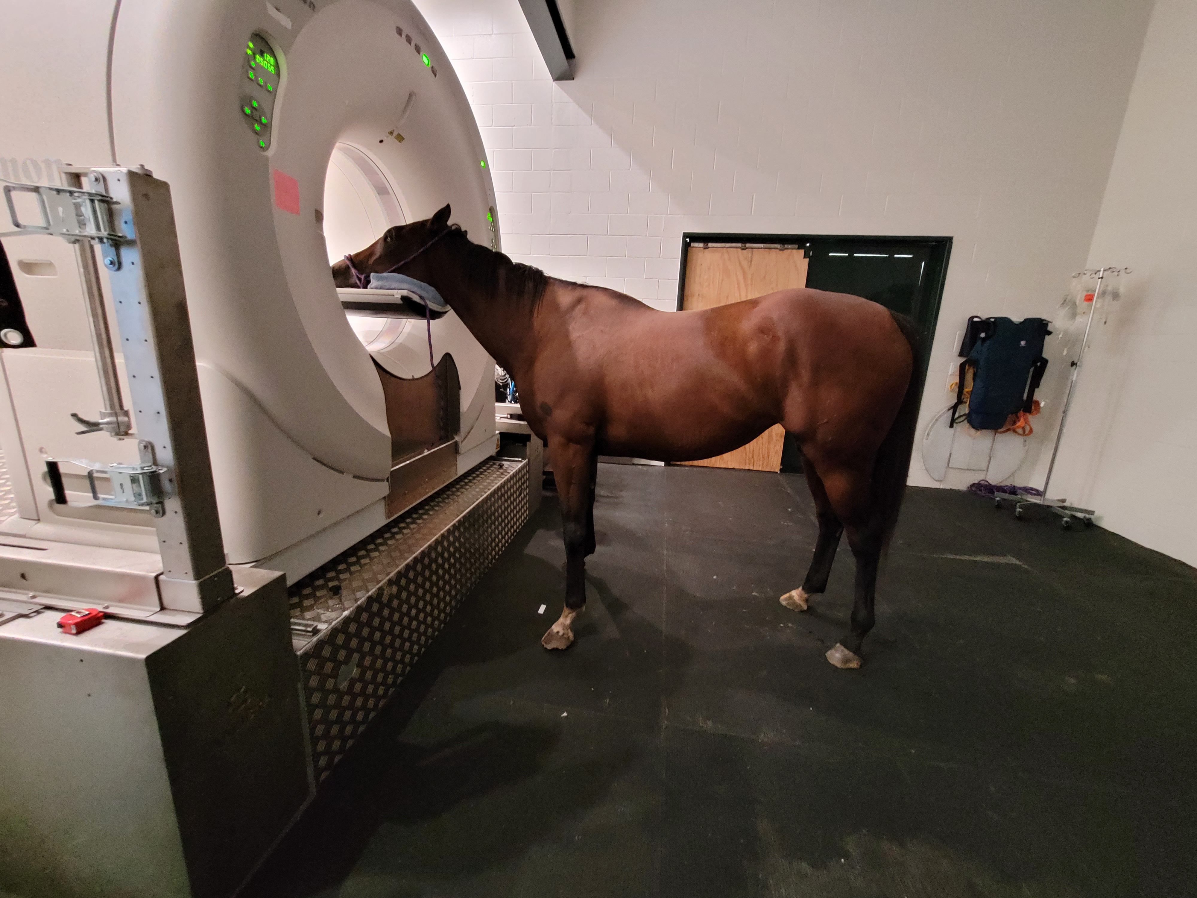
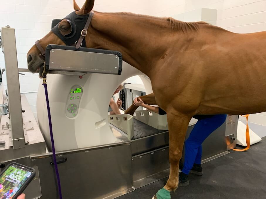
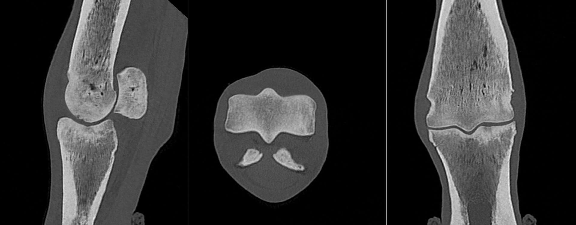
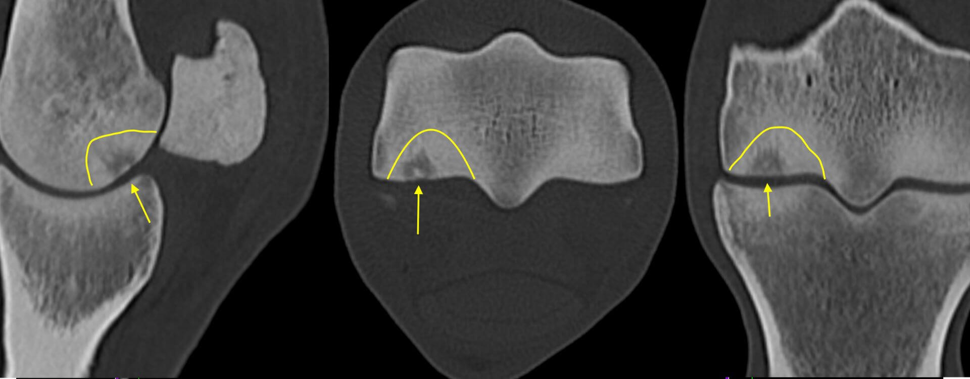
Ocala Equine Hospital installed the first Qalibra CT scanner in North America. This scanner uses a large-bore Canon human CT scanner which has been modified so that the gantry moves over the horse rather than moving the horse through the gantry. The image quality produced is outstanding and any portion of the horse's anatomy may be imaged including shoulders and stifles. Scans of the distal extremities from above the knee or hock to the foot and the head and cranial cervical spine can be done standing with sedation. Scans of the lower neck and upper limbs require a short general anesthesia. Scans of the extremities are typically done with slice thicknesses of 0.5 mm resulting in extremely detailed images. Contrast studies (myelograms, arthrograms, IV contrast studies, etc) can also be done when indicated.