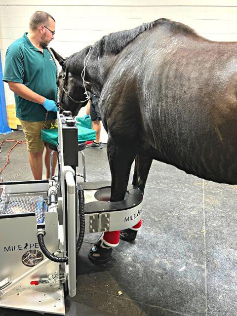
POSITRON EMISSION TOMOGRAPHY (PET scan) is a useful diagnostic tool in pinpointing the source of lameness and it is also very helpful in assessing the healing of boney injuries and some soft tissue injuries answering the question “is my horse ready to return to training?” The PET scanner is able to obtain images from just above the knee or hock to the foot.
How do PET scans work?
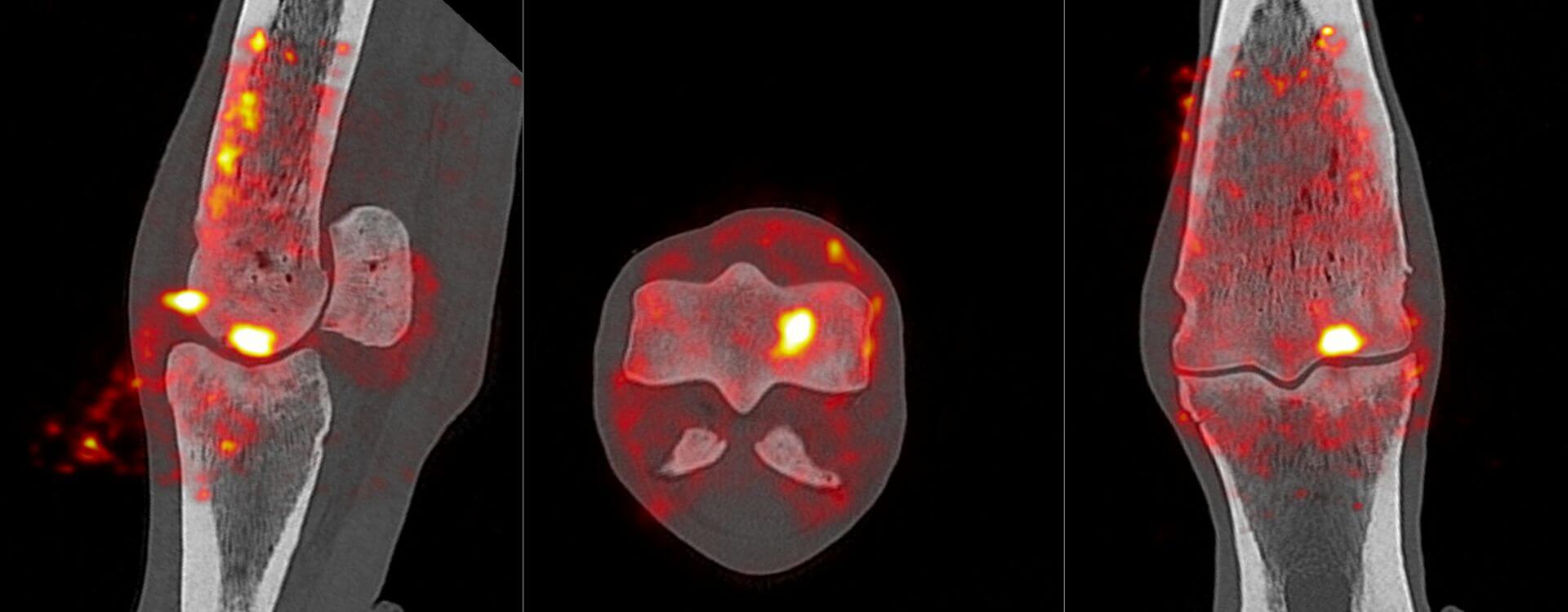
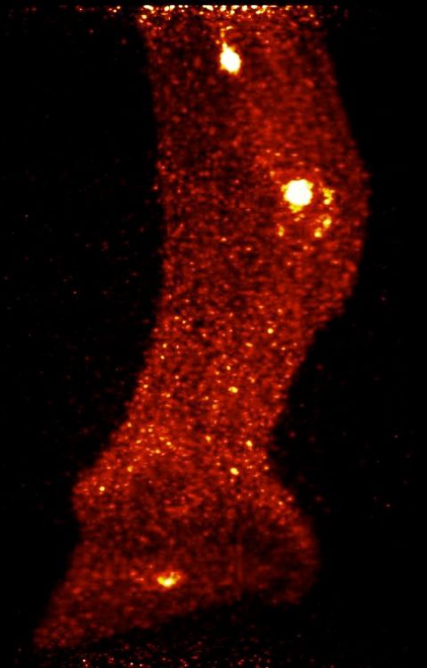
 PET scanning is another functional imaging technique like scintigraphy, however, PET scans are also a volumetric imaging technique that produces images in three dimensions (as opposed to the two-dimensional images of a bone scan).
For a PET scan, a small amount of tracer compound (NaF-18 for bone or F-18 fluorodeoxyglucose for soft tissue) is injected IV and the horse is scanned about 1 hour later. The F-18 emits positrons and the emitted positrons encounter electrons in the adjacent tissue resulting in the release of two gamma-ray photons. As opposed to a flat gamma camera like the device used for a bone scan, the part to be imaged is placed in a ring of detectors analogous to the gantry on a CT device. The NaF binds to areas of increased bone turnover and the FDG is taken up by metabolically active cells at the site of a soft tissue injury. NaF imaging is also exquisitely sensitive for detecting areas of increased bone turnover and the resulting images have far greater anatomical resolution than a two-dimensional bone scan.